Occlusive Disease: Aorta & Iliac Arteries
Occlusive Disease: Aorta & Iliac Arteries
Essentials of Diagnosis
- Cramping; pain or tiredness in the calf, leg, or hip while walking (claudication).
- Diminished femoral pulses.
- Tissue loss (ulceration, gangrene) unusual.
General Considerations
The classic patient with aortoiliac disease is a 50- to
60-year-old male smoker in whom this is the initial manifestation of systemic
atherosclerosis. Occlusive disease of the aorta and iliac arteries begins most
frequently at the bifurcation of the aorta in the proximal common iliac
arteries. Lesions affecting the external iliac arteries are less common. Disease
progression may lead to complete occlusion of one or both common iliac arteries,
which can precipitate occlusion of the entire abdominal aorta to the level of
the renal arteries. Pathologic changes of atherosclerosis may be diffuse, but
flow-limiting stenoses occur segmentally. This is particularly true of the
aortoiliac segment where patients may have limited or no narrowing of the
vessels in the more distal vessels.
Clinical Findings
Symptoms and Signs
Intermittent claudication occurs as a cramping pain brought
on by exercise usually located in the calf muscles. The pain may extend into the
thigh and buttocks with continued exercise. It may be unilateral or bilateral.
Some patients complain only of weakness in the legs when walking, or simply
extreme limb fatigue. The symptoms are relieved with rest. With bilateral
symptoms, impotence is a common complaint. Femoral pulses are absent or very
weak as are the distal pulses. A bruit may be heard over the aorta, iliac, and
femoral arteries.
Doppler Findings
The ratio of systolic blood pressure at the ankle compared
with the brachial artery of the upper arm, as detected by Doppler examination,
is reduced to below 0.9 (normal ratio is 1.0–1.2); this difference is
exaggerated by exercise. Segmental wave-forms or pulse volume recordings
obtained by strain gaze technology through blood pressure cuffs demonstrate
blunting of the arterial inflow throughout the leg.
Imaging
CT angiography (CTA) and magnetic resonance angiography
(MRA) have largely replaced traditional invasive angiography to determine the
anatomic locations of the occlusions. Imaging is only required when intervention
is contemplated, since a segmental wave-form analysis should appropriately
identify the involved levels of the arterial tree.
Treatment
Conservative Care
A program that includes smoking cessation, risk factor
reduction, weight loss, and walking will substantially improve exercise
tolerance. A trial of phosphodiesterase inhibitors, such as cilostazol 100 mg
orally twice a day, may be beneficial in approximately two-thirds of patients.
In the initial stages of a rehabilitation program, substantial benefit may be
derived from simply slowing the cadence of walking.
Surgical Intervention
Intervention to relieve the obstruction is to be considered
if claudication interferes appreciably with the patient's essential activities
or employment. An aorto-femoral bypass graft that bypasses the occluded segments
of the aortoiliac system is a highly effective and durable treatment for this
disease. This prosthetic graft extends from the infrarenal abdominal aorta to
the common femoral arteries. High-risk patients may also be treated with a graft
from the axillary artery to the femoral arteries (axillo-femoral bypass graft)
or, in the unusual case of iliac disease limited to one side, a graft from the
contralateral femoral artery (fem-fem bypass). The less extensive operations
have lower operative risk; however, they are less durable.
Endovascular Techniques
Because the flow-limiting stenoses tend to be segmental,
occlusive lesions of the aortoiliac segment can be effectively treated with
angioplasty and stenting. This approach matches the results of surgery for
single stenoses but both effectiveness and durability decreases with multiple
stenoses.
Complications
The complications of the aorto-femoral bypass are those of
any major abdominal reconstruction in a patient population that has a high
penetrance of cardiovascular disease. Mortality should be low, in the range of
2–3%, but morbidity is higher with a 5–10% rate of myocardial infarction. The
total complication rate may be over 10%. Complications of endovascular repair
include embolization and vessel dissection. These are relatively uncommon and
the total complication rate should be below 5% with this technique.
Prognosis
Without intervention the prognosis for patients with
aorto-iliac disease include reduction in walking distance but rarely include
rest pain or threatened limb loss. Life expectancy is related to their attendant
cardiac disease with a mortality rate of 25–40% at 5 years.
Symptomatic relief is generally excellent after
intervention. After aorto-femoral bypass, a patency rate of 90% at 5 years is
common. Patency rates and symptom relief for less extensive procedures are also
good with 20–30% symptom return at 3 years.
Occlusive Disease: Femoral & Popliteal Arteries
Essentials of Diagnosis
- Cramping; pain or tiredness in the calf only with exercise.
- Reduced popliteal or pedal pulses.
- Foot pain at rest, relieved by dependency.
- Foot gangrene or ulceration.
General Considerations
The superficial femoral artery (SFA) is the artery most
commonly occluded by atherosclerosis. The lesions frequently occur where the SFA
passes through the abductor magnis tendon in the distal thigh. The profunda
femoris artery and the popliteal artery are relatively spared occlusive lesions
except in diabetics. As with atherosclerosis of the aorto-iliac segment, these
lesions are closely associated with a history of smoking.
Clinical Findings
Symptoms and Signs
Symptoms of intermittent claudication are confined to the
calf. Occlusion of the SFA at the abductor canal when the patient has good
collaterals from the profunda femoris will cause claudication at approximately
2–4 blocks. However, with concomitant disease of the profunda femoris or the
popliteal artery, much shorter distances may trigger symptoms. With
short-distance claudication, dependent rubor of the foot with blanching on
elevation may be present. Chronic low blood flow states will also cause atrophic
changes in the lower leg and foot with loss of hair, thinning of the skin and
subcutaneous tissues, and disuse atrophy of the muscles. As the common femoral
artery is rarely affected with occlusive disease, the common femoral pulsation
is usually of good quality, but the popliteal and pedal pulses are reduced.
Laboratory Findings
The ankle-brachial index (ABI) is reduced; levels below 0.5
suggest severe reduction in flow. ABI readings depend on arterial compression.
Since the vessels may be calcified in diabetic patients and the elderly, ABIs
can be misleading and must be accompanied by a wave-form analysis. Pulse volume
recordings with cuffs placed at the high thigh, mid thigh, calf, and ankle will
delineate the levels of obstruction with reduced pressures and blunted
wave-forms. Angiography, CTA, or MRA all adequately show the anatomic location
of the obstructive lesions. Generally, these studies are only done if
revascularization is planned.
Treatment
Conservative Care
As with aorto-iliac disease, conservative management has an
important role for some patients, particularly those individuals with SFA
occlusion and good profunda femoris collaterals. For these patients conservative
management as noted above can result in excellent outcomes with no intervention
required.
Surgical Intervention
Bypass Surgery
Surgery is indicated if intermittent claudication is
progressive, incapacitating or interferes significantly with essential daily
activities. Intervention is mandatory if there is rest pain or threatened tissue
loss of the foot. The most effective and durable treatment for lesions of the
SFA is a femoral-popliteal bypass with autogenous saphenous vein. Synthetic
material, polytetrafluoroethylene (PTFE) can be done with relatively short
bypasses with excellent distal vessels. These grafts do not have the patency of
vein but may have value in patients who have no vein available.
Thromboendarterectomy
Removal of the atherosclerotic plaque is now limited to the
lesions of the common femoral and profunda femoris artery where bypass grafts
and endovascular techniques have no role.
Endovascular Surgery
Endovascular techniques have increased in popularity for
lesions of the SFA. There are several alternatives. Angioplasty may be combined
with stenting either with a bare metal stent or a PTFE-covered stent to form an
endoluminal bypass. Cyroplasty, angioplasty with balloon cooled to a –20° and
endoluminal atherectomy also have their proponents. These techniques have lower
morbidity than bypass but also have a lower rate of success and durability.
The most favorable lesions for endovascular therapy are
lesions that are less than 10 cm long and in patients who are undergoing
aggressive risk factor modification. After any of these procedures, the patient
generally receives lifelong antiplatelet medication, with periprocedural
treatment with clopidogrel (75 mg/day) and long-term maintenance therapy with
aspirin.
Complications
Open surgical procedures of the lower extremity,
particularly long bypasses with vein harvest, have a risk of wound infection
that is higher than in other areas of the body. Leg infection or seroma can
occur in as many as 15–20% of cases. Myocardial infarction rates after open
surgery are 5–10%, with a 1–4% mortality rate. Complication rates of
endovascular therapy are 1–5%, making these therapies attractive despite their
lower durability.
Prognosis
The prognosis for motivated patients with isolated SFA
disease is excellent, and surgery is not recommended for mild or moderate
claudication in these patients. However, when claudication significantly limits
daily activity and undermines quality of life as well as overall cardiovascular
health, intervention may be warranted. All interventions require close
postprocedure follow-up with ultrasound surveillance so that any recurrent
narrowing can be treated promptly to prevent complete occlusion. The patency
rate of bypass grafts of the femoral artery, SFA, and popliteal artery may be as
high as 70% at 3 years with patency for endovascular procedures somewhat
lower.
Occlusive Disease: Lower Leg & Foot Arteries
Essentials of Diagnosis
- Rest pain of the forefoot relieved by dependency.
- Pain or numbness of the foot with walking.
- Ulceration or gangrene of the foot or toes.
- Pallor when the foot is elevated.
General Considerations
Occlusive processes of the lower leg and foot primarily
involve the tibial vessels with only extensive disease involving the arteries of
the foot. There often is extensive calcification of the vessel wall. This
distribution of atherosclerosis is primarily seen in patients with diabetes
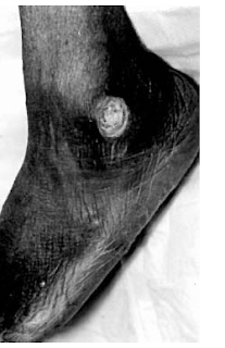
mellitus (see illustration).
Clinical Findings
Symptoms and Signs
Unless there are associated lesions in the aorto-iliac or
femoral/SFA segments, claudication may not be evident. The gastrocnemius and
soleus muscles may receive adequate blood supply from collateral vessels from
the popliteal artery; therefore, when disease is isolated to the tibial vessels,
there may be foot ischemia without attendant claudication, and rest pain may be
the first sign of severe vascular insufficiency. Classically, rest pain is
confined to the dorsum of the foot at the area of the metatarsal heads and is
relieved with dependency. Because of the high incidence of neuropathy in these
patients, it is important to differentiate rest pain from neuropathic
dysesthesia. If the pain is relieved by simply dangling the foot over the edge
of the bed, which increases blood flow to the foot, then the rest pain is due to
vascular insufficiency. The pain is severe, usually burning in character and
will awaken the patient from sleep. On examination, depending on whether
associated proximal disease is present, there may or may not be femoral and
popliteal pulses, but the pedal pulses will be absent. Dependent rubor may be
prominent with pallor on elevation. The skin of the foot is generally cool,
atrophic, and hairless.
Laboratory Findings
The ABI may be quite low (in the range of 0.3), but ABIs
may be falsely elevated because of the noncompressability of the calcified
tibial vessels. Wave-form analysis is important in these patients with a
monophasic flow pattern denoting critically low flow. Segmental pulse volume
recordings will show a fall-off in blood pressure between the calf and ankle,
although pulse volume recordings also may also be affected by tibial vessel
calcification.
Imaging
CTA, MRA, or angiography, is often needed to delineate the
anatomy of the tibial-popliteal segment.
Treatment
Good foot care may avoid ulceration, and most diabetic
patients will do well with a conservative regimen. However, if ulcerations
appear and there is no significant healing within 2–3 weeks, revascularization
will be required (see photograph). Infrequent rest pain is not an absolute
indication for revascularization. However, rest pain occurring nightly with
monophasic wave forms requires revascularization to prevent threatened tissue
loss.
Bypass and Endovascular Techniques
Bypass with vein to the distal tibial arteries or foot has
been shown to be an effective mechanism to treat rest pain and heal gangrene or
ischemic ulcerations of the foot. Because the foot often has relative sparing of
vascular disease, these bypasses have had good patency rates (70% at 3 years).
Fortunately, in nearly all series, limb salvage rates are much higher than
patency rates.
Endovascular techniques are beginning to be used in the
tibial vessels with modest results, but bypass grafting remains the primary
technique of revascularization.
Amputation
Patients with rest pain and tissue loss are at high risk
for amputation, particularly if revascularization cannot be done, or it may be
necessary to debride necrotic or severely infected tissue. Toe amputations, even
of the first toe, have little or no affect on the mechanics of walking. A
transmetatarsal amputation, removing all toes and the heads of the metatarsals,
is durable but increases the energy required of walking by 5–10%. Unfortunately,
the next level that can be successfully used for a prosthesis is at the below
knee level. The energy expenditure of walking is then increased by 50%. With an
above knee amputation the energy required to ambulate may be increased as much
as 100%. While there are good prosthetic alternatives for these patients,
activity levels are limited after amputation, and there are issues relating to
self-image. These factors combine to demand revascularization whenever possible
to preserve the limb.
Recosurces : Current Medical Diagnosis &
Treatment 2008
Stephen J. McPhee, Maxine A. Papadakis, and Lawrence M. Tierney, Jr., Eds.
Ralph Gonzales, Roni Zeiger, Online Eds.
Stephen J. McPhee, Maxine A. Papadakis, and Lawrence M. Tierney, Jr., Eds.
Ralph Gonzales, Roni Zeiger, Online Eds.


